Conditions

Explanation of different Conditions and their Treatments

Welcome to our comprehensive guide on eye care and treatments available in Pantheo.

Below, you’ll find detailed information about common eye conditions and the various treatments offered. At the end of each section, you can download an information booklet in PDF format for your reference.

How The Eye Works

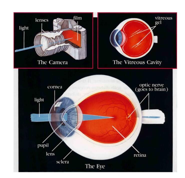
The eye is like a camera. When you take a picture, the lens in the front of the camera allows light through and focuses that light on the film that covers the back inside wall of the camera. When the light hits the film, a picture is taken. The eye works in much the same way. The front parts of the eye (the cornea, pupil, and lens) are clear and allow light to pass through. The light also passes through the large space in the centre of the eye called the vitreous cavity.

The vitreous cavity is filled with a clear, jelly-like substance called the vitreous or vitreous gel. The light is focused by the cornea and the lens onto a thin layer of tissue called the retina, which covers the back inside wall of the eye. The retina is like the film in a camera. It is the seeing tissue of the eye. When the focused light hits the retina, a picture is taken. Messages about this picture are sent to the brain through the optic nerve.

This is how we see.

LenSx Laser Cataract Surgery

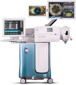
The LenSx laser is an image-guided, high-repetition-rate femtosecond laser that performs the most crucial steps of the cataract surgery with unparalleled precision. It represents the latest development in cataract surgery which is called FLACS (Femto Laser Assisted Cataract Surgery).

For decades, cataract surgeons used manual techniques to perform the intricacies of cataract surgery. Today, surgeons in state of the art Eye Centres around the world have the ability to perform these steps with the Femto Laser.

The LenSx completes the most difficult parts of the surgery with extreme accuracy.  The surgeon using detailed computerized images and advanced OCT imaging of the eye, adjusts the Laser to create precisely positioned incisions in the cornea.  The cataract is then fragmented by the Laser before finally being removed by the surgeon using ultrasound.

LenSx offers several surgical advantages over manual cataract surgery:

  • Precision, control and reproducibility of incisions unachievable with manual techniques.
  • Accuracy in depth and positioning of corneal incisions – critical in astigmatic control.
  • Perfect centration and sizing of the capsulorhexis (opening in the cataract capsule) – critical in optimizing premium multifocal lens implantation
  • Fragmentation of the lens substantially reducing ultrasonic energy use

It is important to understand that phacoemulsification is still a perfectly safe and successful method of removing your cataract.  It remains in fact the most commonly used method.

LenSx gives advantages over phacoemulsification that relate to the accuracy of the final refractive result of the surgery and the need to wear glasses for distance and reading rather than safety of the final visual result.

Preparation instructions for Laser Cataract Surgery

Your cataract will be removed using LenSx Laser Surgery.  This is a completely painless procedure of short duration.  A foldable lens implant is then inserted into the eye and usually sutures are not required.  You will be able to go home immediately after the operation is completed.

LenSx has a very high success rate but like all surgical procedures there is a small risk of complications.

These include vitreous loss, infection (endophthalmitis), choroidal haemorrhage and retinal detachment.  In order to minimise the risk of complicationsit is very important to follow your pre and postoperative instructions carefully.

Pre operatively

  • On the day of surgery have a light breakfastand if you have been prescribed drops to use prior to surgery do so as instructed.
  • If you are takingdiabetic medication, heart or blood pressure tablets take them normally
  • If you are using blood thinning medication or aspirin inform your doctor
  • Report any allergies or previous adverse reactions to procedures or medications.
  • You can come dressed in your normal clothing but short sleeves are preferable so that blood pressure monitoring is not hindered.

On arrival

  • On arriving at the clinic please register with reception and you will be escorted from there to your room.
  • You will be taken to the operating theatre by the nursing staff when it is time for your surgery, this is usually between 1 to 3 hours after your arrival depending on the preparation time necessary and how busy the theatre timetable is on that day.
  • In the operating theatre you will receive some local anaesthetic drops 5 to 10 minutes before your surgery.
  • Usually an injectable local anaesthetic is not necessary, if it is this will be explained to you.
  • During the operation you will be wide-awake and you will be able to hear everything but you will not see or feel any part of the operation. You will be able to talk to the surgeon at any time and tell him if you have a problem.
  • At the end of the procedure your eye will be closed with an eye pad and you will go back to your room.

Post operatively

  • During your first evening after the surgery feel free to do as you please as long as you keep your eye pad dry and intact. If you develop a headache you can take your normal pain relief that you would use at any other time.
  • Your eye pad will either be removed the day of your operation or the following day depending on your case. Your eye will remain open from then on.  You will have reasonably good vision from day one apart from mild haze due to the enlargement of the pupil.  This will improve over a few daysand vision will continue to improve for a couple of months.

LenSx Laser Cataract Booklet

You can download our English “LenSx Laser Cataract” Booklet here

You can download our Greek “LenSx Laser Cataract” Booklet here

Cataract Consultants

Cataract

Dr. Theodoros Potamitis
Consultant Ophthalmologist (CCST UK) and Associate Professor in Ophthalmology St George's University of London Medical School at the University of Nicosia
Trauma, Vitreo-retinal surgery, Medical retina
Dr. Pantelis Ellinas
Consultant Ophthalmologist
Cataract and Refractive Surgery
Dr. Maria Drousiotou
Consultant Ophthalmologist
Cataract and Refractive Surgery
Dr. Katia Papastavrou
Consultant Ophthalmologist
Glaucoma and glaucoma surgery
Dr. EleniTopouzi
Consultant Ophthalmologist
Oculoplastics, lacrimal surgery, cataract
Dr. Yianna Antoniou
Consultant Ophthalmologist
Cataract, Vitreo-retinal surgery, Medical retina
Dr. Michalis Palos
Consultant Ophthalmologist
Cornea and external eye disease
Dr. Antigoni Koukkouli
Consultant Ophthalmologist
Oculoplastics, Thyroid eye disease, Lacrimal surgery and Neurophthalmology
Dr. Loukiana Tsierkezou
Consultant Ophthalmologist
Cataract, Medical Retina and Uveitis (Inflammatory eye diseases)

Cataract

Cataract occurs when the lens of the eye becomes cloudy or opaque it no longer allows light to enter the eye and the vision deteriorates. An opaque or cloudy lens is called a cataract. This is what needs to be removed and replaced in order to restore vision.

Preparation Instructions for Cataract Surgery

Your cataract will be removed using phacoemulsification. This is a completely painless procedure of short duration. It uses ultrasound to liquefy and aspirate the cataract. A foldable lens implant is then inserted into the eye and usually sutures are not required.

You will be able to go home immediately after the operation is completed. Phacoemulsification is the commonest used method of cataract extraction worldwide. It has a very high success rate but like all surgical procedures there is a small risk of complications. These include vitreous loss, infection (endophthalmitis), choroidal haemorrhage and retinal detachment. In order to minimise the risk of complications

It is very important to follow your pre and post operative instructions carefully.

Pre Operatively

  • On the day of surgery have a light breakfast and if you have been prescribed drops to use prior to surgery do so as instructed. · If you are taking diabetic medication, heart or blood pressure tablets take them normally
  • If you are using blood thinning medication or aspirin inform your doctor
  • Report any allergies or previous adverse reactions to procedures or medications.
  • You can come dressed in your normal clothing but short sleeves are preferable so that blood pressure monitoring is not hindered.

Post Operatively

  • During your first evening after the surgery feel free to do as you please as long as you keep your eye pad dry and intact. If you develop a headache you can take your normal pain relief that you would use at any other time.
  • Your eye pad will either be removed the day of your operation or the following day depending on your case. Your eye will remain open from then on. You will have reasonably good vision from day one apart from mild haze due to the enlargement of the pupil. This will improve over a few days and vision will continue to improve for a couple of months.

Cataract Booklet

You can download our Cataract Booklet in English and in Greek

  • Cataract Booklet in English press here
  • Cataract Booklet in Greece press here

Cataract Consultants

Dr. Theodoros Potamitis
Consultant Ophthalmologist (CCST UK) and Associate Professor in Ophthalmology St George's University of London Medical School at the University of Nicosia
Trauma, Vitreo-retinal surgery, Medical retina
Dr. Pantelis Ellinas
Consultant Ophthalmologist
Cataract and Refractive Surgery
Dr. Maria Drousiotou
Consultant Ophthalmologist
Cataract and Refractive Surgery
Dr. Katia Papastavrou
Consultant Ophthalmologist
Glaucoma and glaucoma surgery
Dr. EleniTopouzi
Consultant Ophthalmologist
Oculoplastics, lacrimal surgery, cataract
Dr. Michalis Palos
Consultant Ophthalmologist
Cornea and external eye disease
Dr. Antigoni Koukkouli
Consultant Ophthalmologist
Oculoplastics, Thyroid eye disease, Lacrimal surgery and Neurophthalmology
Dr. Loukiana Tsierkezou
Consultant Ophthalmologist
Cataract, Medical Retina and Uveitis (Inflammatory eye diseases)
Dr Antonis Ioannides
Consultant Ophthalmologist
Refractive Surgery, Cataract Surgery Specialist
Petros Aristodemou
Consultant Ophthalmologist
Specialist in diseases of the Retina, Macula and Cataract

Diabetic Retinopathy

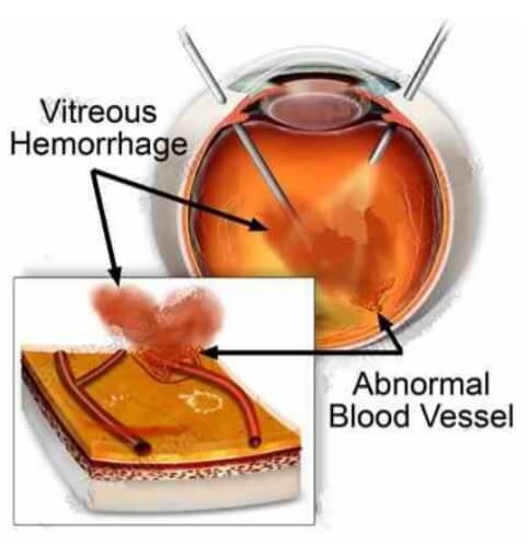
In diabetic retinopathy the blood vessels of the retina become abnormal and develop tiny leaks. These leaks cause fluid or blood to seep into the retina. The retina then becomes wet and swollen (this is called oedema) and cannot work properly. Another problem with the retinal blood vessels in diabetes is that they can close. The retinal tissue, which depends on those vessels for nutrition, will no longer work properly. The areas of the retina in which the blood vessels have closed then trigger the growth of abnormal new blood vessels (this is called neovascularisation) that can be very bad for the eye because they can cause bleeding and scar tissue that can result in blindness.

Fluorescein Angiography and OCT

If your doctor diagnoses diabetic retinopathy and feels that laser surgery might be helpful, a special test called fluorescein angiography may be done. To do the test, dye is injected into a vein in the patient’s arm. The dye travels throughout the body, including the eyes. With a special camera photographs of the retina are taken as the dye passes through them. The photographs will provide a kind of map, which the doctor will use as a guide to the exact location and amount of laser surgery necessary.

OCT is very specialised method of scanning the retina and viewing it in sections. It provides information about the thickness of the macula and which layers of the retina are damaged.

Laser surgery

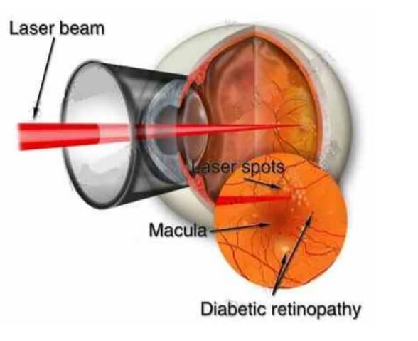
Laser surgery can be very helpful for the treatment of diabetic retinopathy. The laser beam is a high energy light that turns to heat when it is focused on the parts of the retina to be treated. The laser heat seals the leaking blood vessels of the macula and reduces their leakage. Essentially, the major purpose of laser surgery is to prevent further visual loss. Vision is not often improved by laser and it may even be reduced by laser treatment, especially in the initial stages. If however laser treatment is not performed when it becomes necessary then visual deterioration will usually progress rapidly.

Anti VEGF Treatment – Eylea, Lucentis and Avastin

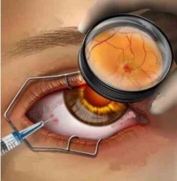
Eylea (aflibercept), Lucentis (ranibizumab) and Avastin (bevacizumab) are anti- vascular endothelial growth factor drugs (antiVEGFs). Injection of an anti-VEGF drug into the vitreous cavity is used to reduce macular oedema (fluid at the back of the eye) and reduce new vessel growth. The injection is administered after the instillation of anaesthetic drops onto the eye which renders the procedure painless. The sterile environment of an operating theatre is used to minimize the risk of infection.

Anti VEGF injections have been shown by studies to improve vision and reduce the progression of diabetic new vessels.

Vitreous Haemorrhage and Vitrectomy

When a person does notice the sudden appearance of floaters, spider webs, spots in front of the eyes, or blurred vision, they should immediately call their eye doctor. Usually these are symptoms of a vitreous haemorrhage. If there is so much vitreous haemorrhage that laser surgery is not possible, or if the blood does not disappear on its own, it can be removed with an operation called a vitrectomy. Vitrectomy surgery is done in the hospital, under general or local anaesthesia. The blood-filled vitreous gel is removed. It is replaced during the operation with a gas bubble or a clear fluid that is compatible with the eye. Over time, the gas bubble or fluid is absorbed by the eye and is replaced by the eye’s own fluid.

Conclusion

Measure your own vision in each eye, separately, each week. Know what you can see in each eye. If you notice a change in your vision, call your eye doctor for an appointment. It is important for all people with diabetes to have a thorough retinal exam regularly, about every six to twelve months, even when there are no problems. Also, be sure to talk to your own medical doctor about the importance of diet and exercise and the dangers of smoking and high blood pressure. And, most of all, learn to maintain the best possible control of your blood sugar. The complications of diabetes, especially diabetic retinopathy, can be reduced by long-term, strict control of blood sugar.

Diabetic Retinopathy Booklet

You can download our Diabetic Retinopathy Booklet in English and in Greek

  • Diabetic Retinopathy Booklet in English press here
  • Diabetic Retinopathy Booklet in Greek press here

Diabetic Retinopathy Consultants

Dr. Theodoros Potamitis
Consultant Ophthalmologist (CCST UK) and Associate Professor in Ophthalmology St George's University of London Medical School at the University of Nicosia
Trauma, Vitreo-retinal surgery, Medical retina
Dr. Yianna Antoniou
Consultant Ophthalmologist
Cataract, Vitreo-retinal surgery, Medical retina
Dr. Loukiana Tsierkezou
Consultant Ophthalmologist
Cataract, Medical Retina and Uveitis (Inflammatory eye diseases)

Droopy Eyelids (Ptosis)

Droopy Eyelids (Ptosis) Consultants

Dr. EleniTopouzi
Consultant Ophthalmologist
Oculoplastics, lacrimal surgery, cataract
Dr. Antigoni Koukkouli
Consultant Ophthalmologist
Oculoplastics, Thyroid eye disease, Lacrimal surgery and Neurophthalmology

Glaucoma

Glaucoma is a disease that damages the optic nerve of your eyes. It usually happens when fluid builds up in the front part of your eye increasing the intraocular pressure. This results to damage of the optic nerve and visual field loss. In most of the cases peripheral (side) vision is affected first, so the patient cannot notice the change. With time, the central (direct) vision will also begin to be lost.

Glaucoma is a leading cause of blindness affecting 80 million people worldwide. Sight loss resulting from glaucoma cannot be reversed. However, early detection and careful, lifelong treatment with medication or surgery can maintain vision.

High intraocular pressure

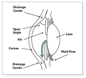
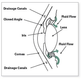
In a healthy eye, a clear fluid, called the aqueous humour is being produced and is essential for the eye to function. It circulates in the front part of the eye and flows out, in order to maintain healthy normal pressure. The outflow is done through a microscopic drain called the trabecular meshwork which is located in the drainage angle.

If the angle is blocked, or does not work properly, then the fluid builds up and the intraocular pressure is raised.
High pressure damages the sensitive optic nerve and results to vision loss.

Optic nerve damage

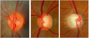
You have millions of nerve fibers that run from your retina to the optic nerve. These fibers meet at the optic disc. As fluid pressure within your eye increases, it damages these sensitive nerve fibers and they begin to die.

As they die, the disc begins to hollow and develops a cupped or curved shape. This causes increase of the cup to disc ratio which is an important measurement to assess the health of your optic nerve. As glaucoma progresses, the cup-to-disc ratio increases. If the pressure remains too high for too long, the extra pressure can damage the optic nerve and result in vision loss.

The average cup-to-disc ratio of a healthy eye is 0.3–0.4. A cup-to-disc ratio of 0.7 or higher is considered an indication of glaucoma. Because early glaucoma typically affects one eye more than the other, a big difference between the cup-to-disc ratios of the two eyes, called asymmetry, is another sign of glaucoma.

During your examination, your doctor will be looking for indications of structural loss of retinal ganglion cells and their fibers in your optic nerve.

This picture shows a healthy optic nerve (left) undergoing the characteristic glaucomatous changes (center) resulting in advanced glaucomatous atrophy (right) and end stage disease.


Visual field loss

Glaucoma is a disease that initially affects your peripheral (side) vision. Often, it is difficult to recognize the loss, because the deficits can be subtle and one eye can compensate for the other.

Visual field testing is performed for every patient to establish the diagnosis of glaucoma and for detecting possible progression.

As the disease progresses, more and more of the peripheral vision is lost until eventually, in very late and advanced disease, the central vision is also affected. Sometimes there are patients with glaucoma who have their central vision affected early in the course of the disease, which is another reason that formal visual field testing is so important.

Over time, visual field testing is performed many times, and while this may seem repetitive and unnecessary, recurrent visual field tests are a critical part of establishing baseline visual fields and monitoring glaucoma over time. This is actually your doctor’s most useful measure of current condition and future risk and also helps determine whether your current treatment is sufficient.

The picture demonstrates the progression of the visual field in a glaucomatous patient if left untreated. Peripheral vision is gradually lost resulting in tunnel vision. In end stage disease even tunnel vision is lost resulting to complete blindness.

Types of glaucoma

There are several types of glaucoma. The two main types are open-angle and angle-closure.

Open-Angle Glaucoma


This is the most common form, accounting for at least 90% of all glaucoma cases:
In these cases the drainage angle is open but not functioning properly, causing increased eye pressure, which leads to optic nerve damage, and possible vision loss.
There are no early warning signs of open-angle glaucoma. It develops slowly and sometimes without noticeable sight loss for many years. Most people who have open-angle glaucoma feel fine and do not notice a change in their vision.


Angle-Closure Glaucoma

In this type, the angle is closed in many or most areas. This rise in eye pressure may occur suddenly (an acute attack of angle closure) or gradually. If the intraocular pressure rises very quickly, it causes symptoms such as eye pain, blurry vision, redness, rainbow colored rings (or haloes) around lights, and nausea and/or vomiting. An acute attack of angle-closure glaucoma can cause permanent vision damage and requires immediate medical attention. The gradual form of angle-closure (chronic angle-closure glaucoma) usually has no symptoms and may not be apparent in the earlier stages without an eye examination.
Treatment of angle-closure glaucoma, and eyes at risk for this disease, usually involves a laser procedure (laser peripheral iridotomy) to create a small opening in the outer edge of the iris.

Detecting glaucoma

In order to detect glaucoma, your doctor will need to perform the following tests:

Tonometry

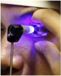
During tonometry, eye drops are intalled to numb the eye and then a device called a tonometer will be used to measure the pressure. The average range for eye pressure is 12–22 mmHg. The level of eye pressure at which glaucoma develops is not the same for everyone and some people can get glaucoma even if their pressures are within the average range of 12–22 mm Hg.


Gonioscopy
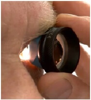
Gonioscopy is a diagnostic exam that helps determine whether the angle formed by the iris and the cornea is open and wide or narrow and closed. During the exam, eye drops are used to numb the eye and a special hand-held contact lens is gently placed on the eye for a few moments.

Pachymetry
Pachymetry measures the thickness of the cornea—the clear window at the front of the eye. Corneal thickness has the potential to influence eye pressure readings. If a cornea is thicker than average, pressure readings with a tonometer may be higher. This gives your eye doctor additional information for your glaucoma diagnosis.

Ophthalmoscopy
This diagnostic procedure helps the doctor examine your optic nerve for glaucoma damage. Eye drops are used to dilate the pupil so that the doctor can see through your eye with a special lens in order to examine the shape and color of the optic nerve. An optic nerve that is cupped or not a healthy pink color is cause for concern.

Perimetry
Perimetry (or a visual field test) produces a map of your field of vision. This test will help your doctor to determine whether your peripheral vision has been affected by glaucoma.

OCT
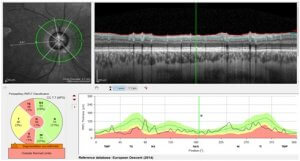
This techonology uses a special laser that produces a three-dimensional high-resolution image of the optic nerve and it measures the thickness of the nerve fiber layer as well as the ganglion cell layer. By imaging your optic nerve over time during multiple visits to your eye doctor, these technologies can help detect progressive loss of optic nerve fibers.

Treatment of glaucoma

With early diagnosis and proper medication and treatment, glaucoma can be controlled. However, sight loss resulting from glaucoma cannot be restored. At the present time, there is no cure. Once detected, glaucoma usually requires ongoing, long-term care. Keeping your eye pressure under control is very important. You must follow your treatment plan carefully to help control your eye pressure. This will protect the optic nerve and prevent sight loss.

Many people think that glaucoma has been cured when high eye pressure is lowered to safe levels with medication or surgery. In fact, the glaucoma is only being controlled, not cured. Regular checkups are still needed even after medications or surgeries have controlled the eye pressure.

Target pressure
The eye specialist will decide which range of pressure is acceptable for each individual case, in order to maintain vision and avoid progression. The target pressure is decided according to the severity of glaucoma, age of patient, corneal thickness, etc.

Glaucoma Medications
Glaucoma is typically treated with the use of medications that either help the fluid drain better or decrease the amount of fluid made by the eye. In most cases, medication can safely control eye pressure for many years.


Laser treatment
There are several types of laser surgery used to treat glaucoma. The type of laser surgery will depend on the form of glaucoma and how severe it is. Laser surgeries are performed in an outpatient setting in your doctor’s office or at the clinic.

Laser surgery is the main initial treatment method for angle-closure glaucoma, which results in widening of the angle for most patients. In cases of open-angle glaucoma, laser surgery can be used as an adjunct treatment with medications.

Incisional Surgery
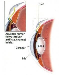
Incisional surgery is usually considered when the maximum amount of medication is not controlling your eye pressure or when your glaucoma is progressing despite normal pressures.

What happens during surgery is that a new drainage is opened for the fluid to drain and the eye to maintain low pressures. The most common types of surgery is trabeculectomy, where the drainage channel is made from the patients own tissues, and tube surgery, where an artificial valve or tube is placed in the eye so that the fluid can drain.
Glaucoma Booklets

You can download our “Understanding Glaucoma” Booklet here

You can download our “Taking Glaucoma Medication” Booklet here

Glaucoma Consultant

Dr. Katia Papastavrou
Consultant Ophthalmologist
Glaucoma and glaucoma surgery

Thyroid Eye Disease

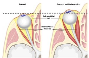
Thyroid eye disease (TED), also known as Graves’ disease, is an autoimmune inflammatory disorder affecting the tissues within the eye socket. It can cause pain, swelling, redness, watering, retraction of the upper eyelids giving a staring appearance, bulging eyes, double vision and occasionally decreased vision.

TED is usually caused by Graves’ disease, a condition that causes over activity of the thyroid gland. However, this condition can be occasionally associated with Hashimoto’s disease or hypothyroidism. TED may occur before or after the onset of systemic hyperthyroid disease and can start suddenly or more slowly. Even though some patients undergo spontaneous remission of symptoms within two years, many need treatment. The active disease course can be prolonged up to 3 years in smokers. Smokers are also at greater risk of sight threatening disease and poorer response to treatment.

The first step of treatment aims to regulate thyroid hormone levels and to stop smoking. Most patients are advised to take Selenium supplements 200mcg once daily for 6 months. Lubricating eye drops are used to avoid damage to the eye. In moderately active disease, signs or symptoms increase and include double vision. Steroids, usually given intravenously, are effective but are used conservatively because of their side effects. Steroids do not reverse the disfigurement caused by the disease but is effective in controlling pain and improving problems with double vision caused by inflamed eye muscles in many cases. In severe cases (3-5%), the patient experiences

intense pain, decrease of vision and loss of colour vision.These occur as a result of sight-threatening corneal ulceration or compression of the optic nerve. This is an emergency requiring immediate treatment.

After the active stage of the disease where the eyes are red and painful the patient may be left with double vision, bulging or staring eyes. These are all amenable to surgery but it may take some time (usually many months to years) for all the treatment to be completed.

TED is a rare but treatable disease that causes a significant decrease in quality of life. Therefore it should be managed with endocrinologists and ophthalmologists working closely together to decide on the best management for you.

Steroid Treatment for Thyroid Eye Disease

You have been recommended to start intravenous steroid treatment for TED due to the significant level of inflammation (moderately active disease) in your eye socket(s) that is causing pain and / or double vision. Steroids are strong drugs and need to be administered in a hospital setting as an infusion into your vein as a day case admission. You are usually admitted under the care of your endocrinologist / thyroid doctor who will be responsible should any systemic complications arise from the treatment. This is a 12-week course of treatment with one infusion given every consecutive week. During the first 6 weeks, you will be receiving 500mg methylprednisolone per session and the following 6 weeks the dose will be decreased to 250mg methylprednisolone.

Intravenous steroids are preferred rather than oral steroid tablets because they are more effective in treating TED and have a better safety profile. Steroids do not reverse the disfigurement (bulging eyes and retracted eyelids) caused by the disease but are effective in controlling pain and improving double vision caused by the inflamed eye muscles.

During the treatment you will need to be monitored in the eye clinic at 2-3 weekly intervals and if you experience double vision you will be seen by an Orthoptist, a specialist technician who measures eye movements. Usually this will take place before / soon after starting your steroid treatment so that we have baseline measurements to monitor your progress with the treatment.

If you experience intense pain, decrease in vision and loss of color vision this is considered an emergency and may require immediate treatment. You will need to contact the eye clinic for an urgent review.

Thyroid Eye Disease Booklet

You can download our Thyroid Eye Disease Booklet in English and in Greek

  • Thyroid Eye Disease Booklet in English press here
  • Thyroid Eye Disease Booklet in Greek press here

Thyroid Consultants

Dr. Antigoni Koukkouli
Consultant Ophthalmologist
Oculoplastics, Thyroid eye disease, Lacrimal surgery and Neurophthalmology
Dr. EleniTopouzi
Consultant Ophthalmologist
Oculoplastics, lacrimal surgery, cataract

Neuro-ophthalmology

Neuro-ophthalmology merges the disciplines of ophthalmology (eyes) and neurology (nervous system), focusing on the complex interaction between the eyes, brain and the nerves. Almost half of our brain is dedicated to the processing of visual images and moving our eyes, therefore it is not surprising there are several conditions that affect these areas.  Neuro-ophthalmologists often spend a significant amount of time with their patients, of whom the majority require long-term follow-up. They have unique abilities to evaluate patients from neurologic, ophthalmologic and medical standpoints to diagnose and treat a wide variety of problems:

  • Optic neuritis
  • Optic atrophy
  • Optic drusen
  • Papilloedema
  • Unequal pupils
  • Migraine
  • Myasthenia gravis
  • Multiple sclerosis
  • Double vision
  • Nystagmus
  • Microvascular cranial nerve palsy
  • Idiopathic intracranial hypertension
  • Pituitary tumours
  • Blepharospasm
  • Photophobia
  • Traumatic brain injury
  • Unexplained visual loss

Here at Pantheo we offer comprehensive clinical care using advanced diagnostic equipment and facilities.  We have a fully integrated ophthalmic group practice and are privileged to be able to offer our patients all the ophthalmic tests necessary for their neuro-ophthalmic assessment in order to give them the best care possible.

  • Orthoptic assessment and Hess charts
  • OCT images of the optic nerve and ganglion cell layer
  • Humphrey visual fields
  • Electrodiagnostics – visual evoked potentials, electroretinogram and electrooculogram
  • Fluorescein angiography
  • Ocular ultrasonography

We work closely with the caring neurologists and neurosurgeons, including the Cyprus Institute of Neurology and Genetics, as to ensure a more multidisciplinary approach to the patient’s care. We invest a lot of time counseling our patients and liaising with their general physician or paediatrician. Finally, as one of the largest ophthalmic centers on the island, we hope in the future to be able to organize patient educational activities and collaborate in clinical research.

Preparing for your neuro-ophthalmology appointment:

  • Bring all your relevant investigations including laboratory tests and reports of MRI and CT scans. These will be scanned and stored into your electronic records and then returned to you.
  • If you have had a CT or MRI performed, please bring a copy of your imaging CD as we often wish to review them ourselves.
  • Bring a complete list of medications with you, including the name and dosage of each medication.
  • You will probably have your pupils dilated during the visit. The eye drops last for 2-4hours and will make things look bright and blurry up close. Please have someone else drive you to the appointment and bring sunglasses.
  • During your appointment you might require further investigations such as Humphrey visual fields, OCT images or orthoptic assessment.

Neuro-ophthalmology Consultant

Dr. Antigoni Koukkouli
Consultant Ophthalmologist
Oculoplastics, Thyroid eye disease, Lacrimal surgery and Neurophthalmology

Keratoconus

Keratoconus is an eye condition in which the normally round dome-shaped clear window of the eye (cornea) progressively thins causing a cone-shaped bulge to develop.

Keratoconus Consultants

Dr. Michalis Palos
Consultant Ophthalmologist
Cornea and external eye disease

Retina Detachment

Retinal detachments often develop in eyes with retinas weakened by a hole or tear. This allows fluid to seep underneath, weakening the attachment so that the retina becomes detached – rather like wallpaper peeling off a damp wall. When detached, the retina cannot compose a clear picture from the incoming rays and vision becomes blurred and dim.

Who is at risk of retinal detachment?

Detachment of the retina is more frequent in middle aged, short sighted people. However, it is quite uncommon and only about one person in ten thousand is affected. It is rare in young adults.

What are the symptoms?

The most common symptom is a shadow spreading across the vision of one eye. You may also experience bright flashes of light and/or showers of dark spots called floaters. These symptoms are never painful. Many people experience flashes or floaters and these are not necessarily a cause for alarm. However, if they are getting severe and seem to be getting worse and you are losing vision, then you should seek medical advice. Prompt treatment can often minimise the damage to your eye.

What is the treatment?

If you get help early, it may only be necessary to have laser or freezing treatment. This is usually performed under a local anaesthetic. Frequently, however, an operation will be needed to repair a hole or put the retina back in place. This is usually done under a general anaesthetic.

In 90 – 95 per cent of cases the retina can be repaired with a single operation. The operation does not usually cause much pain, but your eye will be sore and swollen for a few days afterwards. Typically, you will be hospital for a few hours or an overnight stay, depending on your particular condition. We want to reassure you that the surgeon does not take your eye out of its socket to operate on it.

How much vision can I expect after a successful operation?

This depends on how much the retina has detached and for how long. The shadow caused by the detachment will usually disappear when the retina has been put back in place. If your ability to see fine detail has been damaged before the operation, this may not fully recover afterwards.

What happens after the operation?

You will be encouraged to get up and carry on as usual on the day after the operation, although sometimes you will be asked to keep your head in a particular position to help the healing process. Your eye specialist will prescribe eye drops and you will need to use these for a few weeks.

What happens if the detached retina is not put back in place?

Most people will loose all useful vision if no operation is carried out, or if the treatment is unsuccessful. However, further treatment is usually possible if it does not succeed the first time. Occasionally, if the detachment involves the lower portion of the retina, some vision may recover by itself

Can retinal detachment be prevented?

If your family has a history of retinal detachment, or your doctor finds a weakness in your retina, then preventive laser or freezing treatment may be needed. However, in most cases it is not possible to take preventive action. Retinal detachment does not happen as a result of straining your eyes, bending or heavy lifting.

What about my other eye?

If you have had a retinal detachment in one eye, you are at an increased risk of developing one in the other eye. But there is only about a one in ten chance of this happening.

Retinal Detachment Booklet

You can download our Retinal Detachment Booklet in English and in Greek

  • Retinal Detachment Booklet in English press here
  • Retinal Detachment Booklet in Greece press here

Retinal Detachment Consultant

Dr. Theodoros Potamitis
Consultant Ophthalmologist (CCST UK) and Associate Professor in Ophthalmology St George's University of London Medical School at the University of Nicosia
Trauma, Vitreo-retinal surgery, Medical retina
Dr. Yianna Antoniou
Consultant Ophthalmologist
Cataract, Vitreo-retinal surgery, Medical retina

Age Related Macular Degeneration

Age Related Macular Degeneration (AMD) is a disease that can affect the sharp central vision needed for “straight-ahead” activities like reading, driving, telling time and recognizing faces.

Sometimes AMD advances so slowly that you notice little change in vision.  Sometimes AMD progresses quickly, resulting in rapid vision loss.  AMD causes no pain, but it can rob you of your ability to see what is in front of you.  There are two types of AMD – dry and wet.

Dry AMD

In dry AMD, which makes up about 85% of all AMD cases, light –sensitive cells in the retina die, affecting “straight-ahead” vision.  The most common symptom of dry AMD is slightly blurred vision.  Dry AMD tends to develop slowly, but may develop into a more severe form of AMD called wet AMD.

Wet AMD

In wet AMD abnormal blood vessels grow under the macula.  This may lead to bleeding, scar formation and permanent damage.  Damage occurs more rapidly than in the dry form and tends to lead to more severe loss of central or “straight-ahead” vision.  If spotted in time, treatments for certain forms of wet AMD may reduce or delay vision loss.

Who is at risk

The two greatest risk factors for developing AMD are:

  • Increasing age: About 25% of people over 65 years of age have AMD
  • Having AMD in one eye: Of those with AMD in one eye, about 40% will develop AMD in the other eye within five years.

Other risk factors for developing AMD include:

  • Smoking
  • A family history of the disease
  • Low dietary intake of certain vitamins and minerals
  • Gender – women are at greater risk than men

What are the symptoms

  • Dim, fuzzy or less sharp vision
  • A blind spot in your central vision
  • Seeing objects as wavy or curved
  • Colours that seem washed out and dull

Can I test my vision

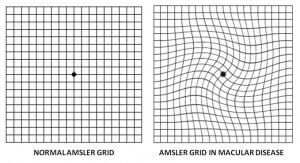
There is no substitute for regular eye examinations with your eye doctor, but there is a way to check for vision symptoms at home.  To test your vision for AMD, use a checkered square known as the Amsler grid.
Use it in good illumination with your reading glasses on at your normal reading distance.  Using one eye at a time focus on the central spot in the grid and observe if there is any distortion on the surrounding lines.  If there is loss of lines or distortion of the lines then this is abnormal.

Ask your eye doctor for an Amsler grid so you can test your vision regularly.  If you detect changes, schedule an eye exam immediately.

Why should I visit an eye doctor?

Regular screening examinations can detect early signs of AMD.  An eye doctor can help you determine your risk for developing AMD, perform an OCT scan to tell you if you have AMD and assist you with treatment options if AMD is diagnosed.  Early diagnosis and treatment may help preserve the sight you still have.

Treatment

There’s currently no cure for either type of age-related macular degeneration (AMD), although the disease process may be slowed down or stopped with the appropriate treatment if used early enough.

Wet AMD

If you are diagnosed with wet AMD the treatment that you will be offered is injections in the eye of a medication called anti vascular endothelial growth factor (anti-VEGF). You may need multiple injections in the eye depending on the response to treatment and in order to achieve stabilisation of vision. The earlier you start the treatment the better the results in gaining vision. Not everyone will see an improvement. The anti-VEGF medications that are usually given are, Aflibercept (Eylea), Ranibizumab (Lucentis) and Avastin (Bevasizumab).

Dry AMD

If you were diagnosed with dry AMD the deterioration of vision is usually slow.  You may go on to develop wet AMD so monitoring is necessary.  There is some evidence a diet high in vitamins A, C and E – as well as substances called lutein and zeaxanthin – may slow the progression of dry AMD, and possibly reduce the risk of getting wet AMD.

Foods high in vitamins A, C and E include:

  • oranges
  • kiwis
  • leafy green vegetables
  • tomatoes
  • carrots

Leafy green vegetables are also a good source of lutein, as are peas, mangoes and sweetcorn.

Dietary supplements are also available if you feel your dietary intake of vitamins is inadequate.

Remember:

  • Timely diagnosis and assessment for treatment of AMD is extremely important
  • Treatment cannot usually bring back vision that is already lost, but the sooner you are treated, the more likely you are to have a better outcome.
  • Your eye doctor can help.

Age Related Macular Degeneration Booklet

You can download our Age Related Macular Degeneration Booklet in English and in Greek

  • Age Related Macular Degeneration Booklet in English press here
  • Age Related Macular Degeneration Booklet in Greek press here

Age Related Macular Degeneration Consultants

Dr. Theodoros Potamitis
Consultant Ophthalmologist (CCST UK) and Associate Professor in Ophthalmology St George's University of London Medical School at the University of Nicosia
Trauma, Vitreo-retinal surgery, Medical retina
Dr. Yianna Antoniou
Consultant Ophthalmologist
Cataract, Vitreo-retinal surgery, Medical retina
Dr. Loukiana Tsierkezou
Consultant Ophthalmologist
Cataract, Medical Retina and Uveitis (Inflammatory eye diseases)

Macular Hole

Macular Hole Consultants

Dr. Theodoros Potamitis
Consultant Ophthalmologist (CCST UK) and Associate Professor in Ophthalmology St George's University of London Medical School at the University of Nicosia
Trauma, Vitreo-retinal surgery, Medical retina
Dr. Yianna Antoniou
Consultant Ophthalmologist
Cataract, Vitreo-retinal surgery, Medical retina

Laser Eye Surgery

Laser Eye Consultant

Dr. Maria Drousiotou
Consultant Ophthalmologist
Cataract and Refractive Surgery
Dr Antonis Ioannides
Consultant Ophthalmologist
Refractive Surgery, Cataract Surgery Specialist

Amniotic Membrane Transplantation

Amniotic membrane is the inner layer of the placenta and it is a thin, avascular tissue which aids healing and inhibits inflammation and scarring.

Indications for amniotic membrane transplantations are as a graft in conjunctival reconstruction; a graft in corneal surface reconstruction; a patch in corneal surface reconstruction; as a substrate in association with limbal transplant in limbal stems cells deficiency (e.g. after an eye burn) and as a barrier in bullous keratopathy in eyes with poor potential (in combination with corneal micropuncturing).

Amniotic Membrane Transplantation Consultant

Dr. Michalis Palos
Consultant Ophthalmologist
Cornea and external eye disease

Uveitis

Uveitis Consultant

Dr. Loukiana Tsierkezou
Consultant Ophthalmologist
Cataract, Medical Retina and Uveitis (Inflammatory eye diseases)